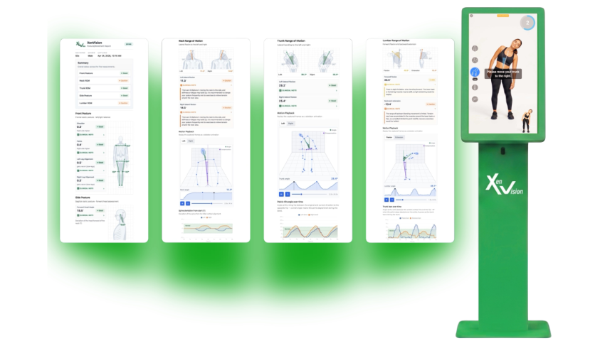
Photo courtesy of Xenco Medical
This article is sponsored by Xenco Medical.
For decades, the fields of computer graphics, computer vision, and orthopedic medicine have evolved along parallel trajectories. SIGGRAPH has continuously advanced the science of image formation, geometric reconstruction, physically based simulation, machine learning, and real-time rendering, while clinical biomechanics has relied upon increasingly sophisticated motion laboratories capable of measuring human locomotion with precision, often requiring expensive infrastructure, reflective markers, force plates, specialized operators, and dedicated laboratory environments.
At SIGGRAPH 2026, Xenco Medical is introducing XenVision, an AI-powered musculoskeletal assessment platform that represents a convergence of these disciplines. Built upon advances in computer vision, geometric modeling, human pose estimation, biomechanical analysis, and medical artificial intelligence, XenVision transforms quantitative movement assessment from a specialized laboratory procedure into a scalable, non-contact screening platform capable of generating assessment results in under two minutes. Rather than replacing traditional gait laboratories or comprehensive orthopedic examinations, XenVision extends the reach of musculoskeletal intelligence by bringing objective functional assessment into orthopedic clinics, employer health programs, hospital systems, and preventive screening environments.
This is not simply another pose estimation application. It is the application of more than three decades of computer vision research toward one of healthcare’s largest unsolved challenges: objective, accessible musculoskeletal screening.
Human movement has long represented one of computer vision’s most technically demanding inverse problems. Unlike rigid scene reconstruction, the human body exhibits articulated kinematic chains, non-rigid deformation, self-occlusion, changing appearance, viewpoint variation, clothing-induced ambiguity, depth uncertainty, and significant inter-subject anatomical variability. Solving these challenges has required foundational advances in projective geometry, camera calibration, robust estimation, feature correspondence, statistical optimization, and multi-view reconstruction, the very mathematical foundations that shaped modern computer vision.
While early motion analysis laboratories relied upon marker-based optical tracking systems and carefully calibrated multi-camera environments, modern deep neural networks have fundamentally changed how anatomical landmarks can be inferred directly from RGB imagery. XenVision leverages these advances to eliminate reflective markers, wearable sensors, and manual anatomical digitization entirely. Sophisticated AI-powered pose estimation algorithms automatically identify anatomical key points, reconstruct functional body geometry, and derive quantitative biomechanical measurements in real time.
At the heart of XenVision is an integrated computational pipeline that bridges computer graphics with clinical biomechanics. The workflow begins with high-fidelity image acquisition optimized for robust skeletal landmark detection. Deep neural pose estimation networks identify anatomical joints while maintaining temporal consistency across entire movement sequences. The resulting skeletal representation becomes the computational substrate for higher-order biomechanical analysis, enabling objective assessment of postural alignment, joint angle estimation, range-of-motion quantification, bilateral symmetry, dynamic functional movement, movement compensation strategies, and longitudinal performance comparison. Unlike consumer fitness applications, XenVision is designed to support quantitative orthopedic assessment. Every inferred landmark becomes part of a mathematically constrained skeletal model from which clinically interpretable biomechanical metrics are derived. The emphasis is not simply determining where joints are located, but understanding how they move through space and how those movement patterns relate to musculoskeletal function.
Modern movement analysis has traditionally depended upon sophisticated biomechanics laboratories equipped with marker-based optical systems, synchronized camera arrays, force platforms, electromyography instrumentation, and highly trained technical personnel. These laboratories remain the gold standard for detailed biomechanical research, yet they are resource intensive and difficult to scale for routine musculoskeletal screening. Biomedical engineering literature has long recognized that objective movement assessment requires sophisticated instrumentation capable of accurately capturing kinematic data while complementing, not replacing, clinical expertise. XenVision approaches this challenge differently. Rather than reproducing every capability of a biomechanics laboratory, it focuses on the portion of musculoskeletal evaluation that benefits most from rapid, repeatable, standardized screening. Its kiosk-based architecture enables completely non-contact assessments without wearable instrumentation, reflective markers, calibration suits, or specialized operator workflows, while automatically generating standardized quantitative reports suitable for clinical interpretation. The result is an assessment workflow measured in minutes rather than hours.
Performance in medical artificial intelligence is determined less by model size than by data quality. XenVision has been engineered using more than 700,000 real-world musculoskeletal assessment datasets, providing a foundation for learning the extraordinary variability present across age, anatomy, pathology, body habits, and functional movement strategies. This large-scale clinical corpus enables the system to recognize subtle deviations in posture and movement that frequently precede symptomatic musculoskeletal disease. Rather than relying solely upon heuristic angle thresholds, XenVision establishes objective functional baselines capable of supporting earlier detection, longitudinal monitoring, and more personalized musculoskeletal health management. The implications are significant. Musculoskeletal dysfunction rarely appears suddenly. Compensation develops gradually, asymmetry accumulates incrementally, and mobility declines progressively over time. Objective quantitative measurement makes these subtle changes visible long before they become clinically obvious through conventional examination alone.
For the SIGGRAPH community, XenVision represents a particularly compelling application of geometric computation. Modern pose estimation is fundamentally a geometric inference problem in which latent anatomical structure must be estimated from projective image measurements while accounting for camera perspective, measurement uncertainty, articulation, temporal coherence, and biological variability. The theoretical foundations originate in decades of research involving projective geometry, camera models, feature correspondence, robust estimation, geometric optimization, multi-view reconstruction, and statistical estimation. These concepts, which have long formed the mathematical backbone of computer vision research, provide the framework for transforming two-dimensional observations into reliable representations of three-dimensional human movement. Although XenVision performs markerless assessment through a streamlined acquisition workflow, its computational architecture reflects the maturation of algorithms originally developed within the geometric vision research community.
XenVision prioritizes communication, including reporting tools intended to help clinicians and patients review assessment results. Following every assessment, patients receive comprehensive digital and printable reports featuring visual movement playback, posture visualization, detailed biomechanical metrics, clinical interpretation, individualized recommendations, and opportunities for longitudinal comparison over time. Complex movement data are transformed into understandable clinical narratives. Clinicians receive standardized quantitative information that supports informed decision-making, while patients receive intuitive visual explanations of their functional movement patterns. This shared language between computational analysis and clinical understanding has the potential to improve patient engagement, treatment adherence, preventive care, and long-term musculoskeletal health management.
Musculoskeletal disorders remain among the leading causes of disability worldwide, yet routine objective movement screening has remained largely inaccessible outside specialized motion laboratories. Computer graphics has spent decades solving difficult problems involving geometry, reconstruction, animation, perception, simulation, and real-time computation. XenVision provides an example of how techniques from computer graphics, computer vision, and biomechanics are being applied in clinical settings. The platform translates innovations from geometric computer vision, deep learning, human motion analysis, AI inference, real-time graphics computation, and biomedical engineering into an orthopedic workflow specifically designed for preventive medicine. In doing so, it exemplifies a broader transformation occurring throughout healthcare: the migration of technologies pioneered within computer graphics and computer vision research into scalable, intelligent clinical systems capable of improving population health.
Throughout SIGGRAPH’s history, the conference has introduced technologies that have ultimately transformed entire industries — from programmable graphics pipelines and GPU computing to physically based rendering, neural graphics, differentiable rendering, and AI-assisted content creation. XenVision represents another example of graphics research extending far beyond visualization and entertainment into clinical medicine. Its significance lies not merely in markerless motion capture, but in demonstrating that advances in geometric artificial intelligence, human pose estimation, and computer vision have matured into practical technologies capable of supporting orthopedic care assessment at population scale.
For the SIGGRAPH community, XenVision serves as a powerful reminder that algorithms originally developed to understand synthetic worlds increasingly possess the fidelity required to understand the human body itself. As artificial intelligence continues to unify computer graphics, computer vision, biomechanics, and healthcare, platforms like XenVision suggest that the next frontier of graphics innovation may not reside solely within virtual environments, but in the quantitative understanding of human movement, functional health, and preventive medicine. In that sense, Xenco Medical’s unveiling of XenVision at SIGGRAPH 2026 is a notable demonstration of how the mathematical foundations of computer graphics and computer vision are beginning to redefine the future of orthopedic care.